
Sternal Elevation
Visualization and dissection across the mediastinum in patients with severe pectus excavatum can be impaired by the inward displaced sternum. Forced sternal elevation increases the anterior-posterior dimension and improves visualization and facilitates mediastinal dissection (Figure 1 a, b,c).


Figure 1a. Intraoperative Photo showing a severe pectus excavatum deformity with significant cardiac compression. Most of the heart is compressed and deviated into the left chest with limited ability to visualize cardiac structures (b) Forced Sternal elevation with a Clamp on the sternum and RulTract retractor has lifted the deformity. (c) There is now clear visualization of the heart borders and adequate space between sternum and pericardium to safely dissect across to the left thorax.
(Photos by permission of DE Jaroszewski)
Sternal elevation is recommended to decrease the risk of pericardial trauma and cardiac perforation. Once the chest has been lifted, rotation and insertion of bars can be performed with reduced stress on the intercostal spaces. This may lessen the risk of intercostal muscle stripping and bar migration.
A variety of techniques can be used to elevate the sternum. In the very young patient with flexible cartilage, a vacuum bell can be applied externally, eliminating the need for any incisions or intrusion upon the sternum on intrathoracic space (Figure 2) (Schier 2005).

Figure 2. Intraoperative application of the vacuum bell in young patients can reduce the pectus defect and allow the sternum to elevate improving visualization and increasing the mediastinal space.
(Photo by permission of RJ Obermeyer)
Wire stitches can be sutured thru the sternum and subsequently attached to a bedside retractor such as the RulTract (RulTract, Inc) or Crane (Primemedkorea). (Yoon 2010, Park 2010). Clamps of varied types can also be placed so that the prongs only enter the anterior table of the sternum and ideally never enter the true thoracic space (Jaroszewski 2013). The clamp is placed either parallel or perpendicular to the sternum in deeper cases to prevent fracture (Figure 3 a, b) and attached to the retractor (Jaroszewski 2013, Park 2010, Belcher 2008).

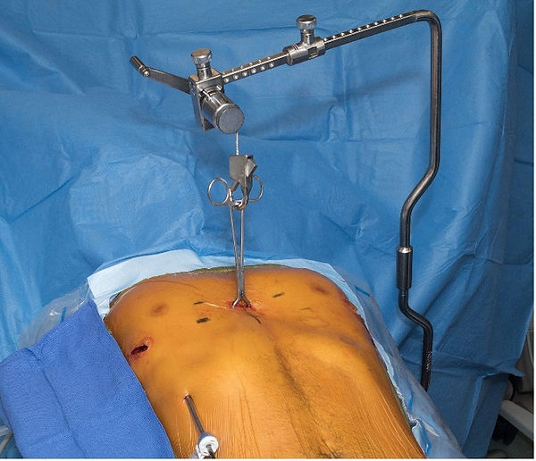
(Photo and drawings by permission of DE Jaroszewski and Mayo Clinic)
Figure 3a,b A Lewin spinal perforating forceps (V. Mueller NL6960, CareFusion, Inc) in inserted into the anterior table of the sternum either perpendicular or parallel to the sternum and attached to the Rultract Retractor (Rultract Inc, Cleveland, OH)
Other authors have reported use of substernal retractors placed either circumferentially or subxiphoid. (Takagi 2012, Tedde 2012, Belcher 2008). Bilateral T-fasteners have also been described with placement in both thorax lateral to the sternum. The sutures are then attached to a Thompson retractor or Rultract.(Kim 2014).
Forced sternal elevation can significantly improve visualization by elevating the defect. It decreases the risk of trauma to the pericardium and the potential for cardiac perforation when dissecting across the mediastinum and placing bars. There is also likely a reduced amount of stress on the intercostal spaces during bar insertion and rotation. We believe this technique can be successfully utilized to improve safety and successful outcomes of minimally invasive pectus excavatum repair.
Author
Dawn Jaroszewski, M.D.
References
Belcher et al. Cardiothorac Surg. 2008
Jaroszewski et al. J Thorac Cardiovasc Surg. 2014
Johnson et al. Ann Thorac Surg. 2013
Kim et al. Ann Thorac Surg. 2014
Park et al. J Thorac Cardiovasc Surg. 2010
Park et al. J Chest Surg. 2021
Schier et al. Ped Surg. 2005
Takagi et al. Pediatr Surg Int. 2012
Tedde et al. Eur J Cardiothorac Surg. 2012
Yoon et al. World J Surg. 2010