
Pectus Excavatum
Pectus Excavatum (PE), the most frequent thoracic malformation, is characterized by a variably deep sternal depression associated to a defect of the condrosternal joints which produces a caved-in thoracic appearance, also known as “funnel chest” .
The condition often involves only the lower part of the sternum (short defect, photo1) but the depression could begin just below the insertion of the second rib (grand canyon defect, photo 2).
PE can be symmetric or asymmetric, with asymmetry associated with sternal rotation to the more depressed side, where the cartilage ribs are shorter.
PE shows a familial recurrence in up to 40% of cases.
PE occurs in an estimated 1 in 300-400 (male to female ratio of 3-5:1) and it is usually noticed shortly after birth, with most cases identified by the first year of life. However, few patients with PE do come to medical attention just before puberty when the condition abruptly accelerates, and a mild defect may quickly turn into a severe one.
PE patients have often a typical aspect: they are slim and tall, with rounded shoulders, a kyphotic habit and a “pot belly” abdomen.
PE can be associated with scoliosis and connective tissue disorders such as
Marfan, Noonan and Ehlers-Danlos syndromes.


Photo 1: Short PE Photo 2: “Grand Canyon” PE
Author
Antonio Messineo, M.D.
References
Nuss et al. Journal Ped. Surgery. 1998
Hebra et al. Thorac. Cardiovasc. Surgery. 2009
Jaroszewski et al. J Am Heart Ass. 2022
Cardiopulmonary Impairment
Considered by some to be a just a cosmetic defect, there is increasing evidence to suggest that pectus excavatum is indeed associated with cardiopulmonary dysfunction. Preoperative cardiac MRI, CPET, and PFT findings in mostly adolescents demonstrated abnormal biventricular systolic function in 16-22% of patients, with most of the remainder having low normal function despite being otherwise young, healthy patients.1, + data by RLB Pectus excavatum was also found to be associated with decreased exercise capacity in 38% and decreased pulmonary function in 23% of these patients.1, + data by RLB Furthermore, the severity of the pectus defect was associated with impaired cardiopulmonary function, with Haller index and correction index being the most useful predictors of impairment.1, 2 Another correlation was demonstrated between severity of pectus deformity (higher Haller index on chest CT) and decreased VO2 max.3 An abnormal VO2 max was also found in 68% of adult patients preoperatively4, while in an adolescent cohort from another study, only 38% of patients had an abnormal VO2 max1, suggesting that exercise capacity decreases significantly with age in patients with pectus excavatum. Pectus excavatum was also found to be associated with decreased right ventricular ejection fraction (RVEF) that increased after repair.5, 6 Abnormal RV systolic function was shown in 16%, abnormal RV diastolic function in 29%, and abnormal septal movement in 69%, all of which were related to pectus severity and worsened with stress.6 A useful classification of patients was published according to site of cardiac compression: Type 0 (no cardiac compression); Type 1 (RV compression); and Type 3 (RV and atrioventricular groove compression) and correlated the site and degree of compression, especially Type 3, with increased stress-related systolic and diastolic dysfunction, septal flattening, and pericardial effusion. 7,8 Age and gender are also significant influencing factors on the cardiopulmonary impact of pectus excavatum. Older patients (18 years and above) are more symptomatic with deeper, more asymmetric defects with diminished exercise capacity and pulmonary function compared to younger patients less than 18 years, and, interestingly, females have better cardiac function, exercise capacity, and pulmonary function than males, despite being more symptomatic and having deeper pectus excavatum deformities.1,9
Despite increasing and compelling evidence to support the negative impact of pectus excavatum on cardiopulmonary function10, the lingering question, especially relevant to payors, is whether pectus repair improves this dysfunction. It has been demonstrated that surgical repair relieved right heart compression and was associated with a significant improvement in right heart chamber size (right atrium, tricuspid annulus, and RV outflow tract) and cardiac output.11 The impact was most striking in adults >30 years who had a >65% increase in RV cardiac output after repair. Further TEE studies also demonstrated improvement in RV global longitudinal strain immediately after repair.12 Consistent improvements in multiple pre- and post- measures of exercise capacity could be demonstrated on CPET (VO2 max, O2 pulse, VO2 max at anaerobic threshold, work, and maximal ventilation) as well as a significant increase in RV stroke volume on TEE at time of bar removal.4 Interestingly, severity of pectus deformity as measured by pectus and cardiac deformity indices (Haller Index, correction Index, sternal tilt, or cardiac compression index) did not correlate with improvements in cardiopulmonary function.4 In a pediatric patient cohort, there were also significant improvements after surgery in exercise capacity (measured by VO2 max, O2 pulse, maximum volume ventilation (MVV), and maximum expired minute ventilation (VE max) as well as forced expiratory volume at 1 second (FEV1)).3
In summary, there is mounting evidence to support that pectus excavatum is not just a cosmetic deformity but is associated with significant cardiopulmonary dysfunction in both pediatric and adult patients that is improved by correction of the deformity, thereby relieving cardiac compression and increasing thoracic volume. Pectus excavatum is associated with low normal to abnormal cardiac function at rest and with decreased exercise capacity that appears to worsen with age. Despite compression of the right side of the heart, the most anterior structure in proximity to sternum, both right and left ventricular function are affected. Both the heart and lungs are affected, although the heart seems to be more impacted than the lungs. Pectus repair is associated with increased subjective improvement in symptomatology and body image as well as objective improvements in cardiac function, exercise capacity, and pulmonary function.
Author
Rebeccah L. Brown, M.D.
References
Zens et a. Ann Thorac Surg. 2022
Abu-Tair et al. Ann Thorac Surg. 2018
Das et al. Ann Pediatr Cardiol. 2019
Jaroszewski et al. J Am Heart Assoc. 2022
Töpper et al. Cardiovasc Thorac Surg. 2016
Raggio et al. Erratum in: Radiol Cardiothorac Imaging. 2020
Rodriguez-Granillo et al. Eur Heart J Cardiovasc Imaging. 2020
Tandon et al. Journal of Cardiovascular Magnetic Resonance. 2014
Casar Berazaluce et al. Pediatr Surg Int. 2020
Tandon et al. Journal of Cardiovascular Magnetic Resonance. 2014
Maagaard et al. Ann Cardiothorac Surg. 2016
Chao et al. Am J Surg. 2015
Chao C et al. Ann Thorac Surg. 2018
Quality of Life
Pectus excavatum often results in a reduced quality of life (QoL) due to a disturbed body image and lower self-esteem. Several studies have shown a significant reduction of QoL compared to controls with normal chestwall. There is no correlation of the severity of the deformity and the reduction of QoL. After correction of the deformity, pectus patients had a significant improvement of QoL.
Author
Caroline Fortmann, M.D.
References
Lomholt et al. Ann Cardiothorac Surg. 2016
de Carvalho et al. J Pediatr Surg. 2020
Kelly et al. Pediatrics. 2008
Minimally Invasive Repair of Pectus Excavatum
(MIRPE)
The minimally invasive repair of pectus excavatum, introduced by Donald Nuss in 1997, represented a complete departure from the previous methods of repair of chest wall deformities. Rather than resection of costal cartilage and sternal fracture, Nuss introduced a corrective procedure. This arose from his recognition that the pliable chest wall of children did not require removal of cartilage is, but rather repositioning the sternum and chest wall.
Briefly, the Nuss procedure involves placing a convex steel bar under the depressed sternum of a patient with pectus excavatum. This is accomplished through small, 2 to 3 cm incisions in the mid axillary line. Using a thoracoscope for visualization, and with the sternum elevated, a plane is created between the front of the pericardium and the back of the sternum. The convex bar, bent to fit the patient’s chest, is passed into the chest medially, then guided through the newly created space behind the sternum across the chest upside down, brought out the other side, and is turned over, immediately pushing the depression of the chest to a normal position. The bar next requires stabilization. This can be accomplished in different ways, including wrapping the bar to the underlying ribs, and utilizing small stabilizing devices on the anterior chest wall subcutaneously.
After allowing approximately three years for the chest to adjust to its new normal position, the bar is removed, usually in an outpatient operation. Removal is accomplished by opening the incisions on both sides and then flattening the bar with an instrument designed for the purpose. Traction on the bar removes it, often with minimal resistance.
The success rate of the operation is extremely high, and complications occur very infrequently in experienced hands.
Author
Robert E. Kelly, Jr. M.D.
Vacuum Bell Therapy
For decades, open surgical repair like the Ravitch procedure was the gold standard to correct Pectus excavatum (PE). The non-surgical technique of applying suction to the ventral chest wall to elevate the sternum was first described in a textbook of Pediatrics in the early 19th century [1], and taken up in the early 20th century by an engineer, using a vacuum bell (VB) made from silicon [2, 3]. An essential paradigm shift in the treatment of PE occurred with the introduction of the technique of minimally invasive repair of Pextus excavatum (MIRPE) by D. Nuss in 1998. The introduction of MIRPE as well as VB therapy (VBT), and an increasing interest and patient’s introspection have changed the view on the treatment of PE within the last 15-20 years. An increasing number of Pediatric PE patients (< 10 years of age) with a mild degree of PE are referred to the outpatient clinic. In many cases of these Pediatric patients, the degree of pectus deformity does not immediately warrant surgery, yet patients may benefit from some type of non-surgical treatment. Furthermore, reports on character and number of possible complications associated with the MIRPE [4] have made conservative VBT a focus of interest of patients, their parents as well as surgeons.
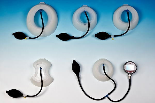
A suction cup is used to create a vacuum at the anterior chest wall. A patient-activated hand pump is used to reduce the pressure up to 15% below atmospheric pressure. Different sizes of vacuum bell exist which are selected according to the individual patients age [figure 1]. When creating the vacuum, the lift of the sternum is obvious and remains for a different time period. The device should be used for a minimum of 30 minutes (2/day), and may be used up to a maximum of several hours daily. Presently a 18-24 month course of treatment is recommended.
Author
Frank-Martin Häcker, M.D.
References
Lange. Pfaundler M, Schlossmann A (editors): Handbuch der Kinderheilkunde, Vol V. Chirurgie und Orthopädie im Kindesalter. Leipzig, FCW Vogel 1910
Bahr. Schwabegger A (editor). Congenital thoracic wall deformities. Berlin-Heidelberg, New York 1st ed. Springer, 2011
Schier et al. J Pediatr Surg 2005
Becmeur et al. J Laparoendosc Adv Surg Tech 2011
Modified Ravitch Technique
The modified Ravitch operation involves resection of all abnormal or deformed costal cartilages, a concept that is equally applicable to excavatum and/or carinatum deformities. Afterwards, it only takes one or two fractures of the anterior table to restore a normal position of the sternum. For cosmetic reasons, consideration should be given to the use of transverse, inframammary incisions, instead of vertical median incision.
After adequate subcutaneous dissection, which begins in the midline and moves laterally, pectoralis muscle flaps are created, exposing the costochondral junction. The defect may involve many cartilages, but usually only cartilages 3 to 7 bilaterally are altered. In most patients, a minimum of four cartilages on each side must be excised. In each of the cartilages involved, the perichondrium is incised longitudinally, exposing the deformed cartilage (Figure 1). Care must be taken to avoid violating the pleural space. Each altered cartilage is resected from the ossified part for sternal fixation. The perichondrium should be preserved as a template for new cartilage growth (Figure 2).

Figure 1: Subperichondrial dissection

Figure 2: Maintainance of the integrity of the perichondrial bundles, to be closed at the end
The xiphoid process is exposed and elevated, and the retrosternal plane is bluntly dissected, moving the pleura and pericardium away from the sternum. The intercostal muscles and perichondrial bundles are partially separated from the sternum, from the xiphoid to the most cranial ribs involved. According to the technique being performed, only the intercostal spaces necessary for fixing the metal bar or mesh should be released, to keep the correction stable and allow closure with a normal conformation of the rib cage.
A single oblique wedge, or transverse osteotomy of the anterior table of the sprained sternum allows for sternal rotation to a neutral position. Occasionally, a second anterior table osteotomy is required. The sternal periosteum is then sutured to keep the sternum in its new, corrected position (Figure 3).

Figure 3: Transverse wedge osteotomy and suture with steel stitches
In young patients, it is highly recommended that you place substernal stainless-steel or titanium bars, or other techniques such as double meshes, under the distal sternum and secure them to the ribs, especially when patients are affected by connective tissue abnormalities. The defect between the sternum and the resected bundles must be closed, bringing these tissues closer together (Figure 4).

Figure 4: Two substernal titanium bars to support the sternum in the correct position
Subsequently, muscular and subcutaneous tissues have to be brought together, ideally using a vacuum suction catheter, with one branch in a subcutaneous position and the other one in a submuscular position.
Post-operative care: The costal cartilages can regenerate 2 to 3 months after the procedure. Therefore, contact sports should be avoided during this period, and some groups recommend at least 6 months for full athletic practice. Patients who have been fitted with steel or titanium bars usually have them removed after 6 to12 months (no later than 12 months, as the fibrosis that sets in can be intense, making it very difficult to identify and remove the bars).
In our opinion, the modified Ravitch procedure should be reserved for adult patients who have a more rigid chest wall and more pronounced defects (deep and asymmetric, with torsion of the sternum), when other procedures are considered inappropriate or risky. Indications may also include patients who have failed a previous Ravitch or modified procedure, or those who had a sternotomy in childhood and developed iatrogenic PE.
Author
José Ribas Milanez de Campos, M.D.
References
Ravitch. J Thorac Surg. 1952
Sulamaa et al. Acta Chir Scand. 1959
Haller et al. Ann Surg. 1976
Mao et al. J Pediatr Surg. 2017